China's Advanced Microscope Enhances Understanding of Mammal Cell Interactions
China's advanced super microscope is enhancing our understanding of how mammal cells interact. The innovative technology allows researchers to delve deeper into cellular processes, leading to new discoveries in biology. By observing these interactions at a much finer resolution, scientists can gain insights that could impact various fields, including medicine and genetics.
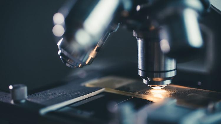
A team of scientists from China has developed an advanced intravital microscope capable of clearly visualizing the intricate three-dimensional interactions within a large-scale cell network at the level of mammalian organs.
This innovative instrument, created by researchers at Tsinghua University, aims to propel forward research in fields such as oncology, immunology, and neuroscience. It seeks to deliver a comprehensive understanding of organ organization and function at the resolution of individual cells.
Known as the RUSH3D system, this microscope provides a centimeter-scale field of view along with subcellular resolution. It can perform high-speed 3D imaging at a remarkable rate of 20 frames per second, while also allowing for prolonged observation of dozens of hours with minimal toxicity, as detailed in a study published on Friday in the journal Cell.
In the realm of neuroscience, the complex interactions among large populations of neurons contribute to higher functions such as intelligence and consciousness. Grasping the structure and operational dynamics of neural circuits is essential for unraveling the brain's inner workings.
The research team employed the RUSH3D system to conduct high-speed 3D observations that examined layers of the cerebral cortex in live mice at single-cell resolution. They were able to capture the various response patterns of distinct cortical regions during multi-sensory stimulation and track extensive neural responses with single-neuron precision over several consecutive days, according to the study.
"The traditional fluorescence microscopy allowed us to observe only part of an organ, such as a specific brain region in a mouse," remarked Dai Qionghai, the paper's corresponding author from Tsinghua University.
"The RUSH3D system, however, is akin to using 100 microscopes simultaneously, providing complete coverage of the mouse cortex and capturing the dynamic interactions of hundreds of thousands of neurons," Dai added.
Frederick R Cook contributed to this report for TROIB News
Discover more Science and Technology news updates in TROIB Sci-Tech